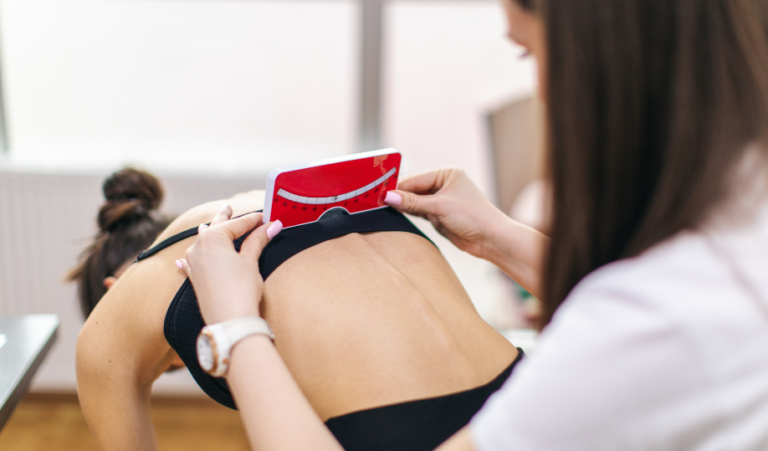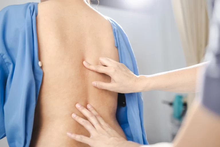
One of the most common concerns I hear from parents at Simply Move Chiropractic is about the safety of repeated X-rays for children diagnosed with scoliosis. Understandably, no parent wants to expose their child to unnecessary radiation. When scoliosis is diagnosed, however, X-rays are the gold standard for monitoring curve progression, especially during rapid growth phases. So how do we balance the need for regular imaging with the desire to protect our children’s long-term health?
To answer this question, I’ve reviewed several key studies examining the effects of repeated spinal X-rays in children and adolescents with scoliosis. The findings are reassuring, and I’d like to share them here to help parents feel confident in the care decisions we make together here in Charlotte, NC.
What the Research Shows About X-ray Exposure
1. The Radiation Risk from Spinal X-rays Is Very Low
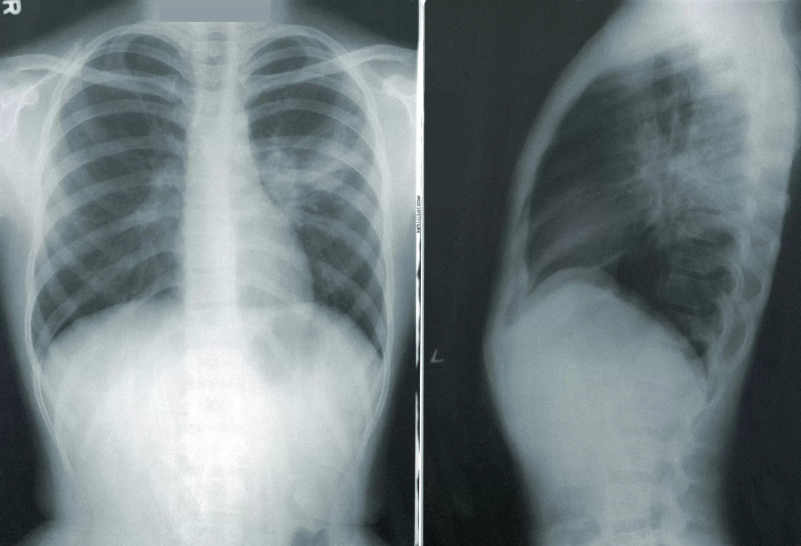
Multiple studies have assessed the actual dose of radiation received during scoliosis X-rays, and the results show it is extremely low—especially with modern digital imaging techniques. According to a review published by Konbaz et al., the average dose per scoliosis film is about 0.1 to 0.5 mSv (millisieverts), which is comparable to the natural background radiation we’re all exposed to in just a few months of daily life.
To put that into perspective, a single chest CT scan may expose someone to 6–7 mSv—more than ten times the dose of a typical scoliosis X-ray. Additionally, today’s X-rays are taken using collimation (to narrow the beam), shielding (like lead aprons), and digital systems that require far less radiation than older film-based methods.
2. No Proven Link Between Scoliosis X-rays and Increased Cancer Risk
One concern some parents raise is whether repeated X-rays could increase cancer risk later in life. A large retrospective study (Oakley et al.) looked specifically at scoliosis patients who had undergone multiple X-rays throughout adolescence. The authors found no statistically significant increase in cancer-related deaths in patients who were monitored with X-rays over many years.
Even in studies where a slightly higher cancer risk was suggested, the increase was very small and difficult to separate from other factors like genetics, hormonal changes, or lifestyle exposures. The vast majority of scoliosis patients, even those who had dozens of X-rays during growth spurts, did not show any clinically meaningful increase in cancer rates.
3. Accurate Imaging Is Essential for Early and Effective Treatment
Another major takeaway from these studies is that early detection and precise monitoring actually reduces the risk of future complications. Without periodic imaging, it’s difficult to know whether a child’s scoliosis is stable, improving, or progressing. In cases where curves progress silently, delayed treatment can lead to more invasive procedures later on—including bracing, surgery, or long-term pain management.
When we have up-to-date imaging, we can respond early with personalized, non-invasive treatment options. At Simply Move Chiropractic, that might include ScoliBrace, ScoliBalance exercise therapy, and postural re-education—all of which are most effective when curve progression is caught early.
4. X-rays Can Now Be Tailored to Minimize Exposure Even Further
Even though I don’t perform imaging in my office, I work closely with local radiology centers and orthopedic providers who specialize in scoliosis imaging. When your child needs an X-ray, there are simple ways to make sure the process is as safe as possible—and as a parent, you have every right to ask about them.
One of the most effective ways to reduce radiation exposure is to request that X-rays be taken using a posteroanterior (PA) view rather than an anteroposterior (AP) view. In a PA view, the X-ray beam enters from the back, which dramatically reduces radiation to sensitive front-facing tissues like the thyroid gland and breast tissue. Research shows that this one change can cut radiation exposure to those areas by up to 90%.
You can also ask whether the imaging center uses thyroid and gonadal shielding during the procedure. These lead shields are designed to protect highly sensitive areas without interfering with the image quality needed to assess scoliosis progression (most of the time). Most pediatric imaging centers already follow these guidelines, but it never hurts to confirm.
Finally, as I mentioned earlier, most modern X-ray machines use significantly lower radiation doses than older models. If you’re still concerned about exposure, you can look for imaging centers that offer EOS imaging—an advanced technology that delivers even lower radiation than standard digital X-ray systems. However, keep in mind, with these systems you need to keep perfectly still for upwards of 15-20 seconds per x-ray. This can be a challenge for young children and may result in the need to repeat the x-rays.
Reassurance for Parents
If you’re a parent navigating a scoliosis diagnosis, I understand how overwhelming it can be. You’re being asked to make decisions about bracing, therapy, and long-term monitoring, all while worrying about the health of your growing child. My goal is to support you with accurate information, compassionate care, and a treatment plan that is both safe and effective.
The research is clear: when done appropriately, scoliosis X-rays are safe and carry minimal risk. They are an essential part of tracking spinal development and ensuring we catch any changes early—when conservative treatment options are most effective.
So yes, your child may need periodic imaging as they grow. But with today’s technology and protocols, those X-rays are a small and worthwhile tool in the journey to better spinal health.